Fabrication of 3D electrode arrays through sidewall-metallization of SU-8 micropillars integrated with open-well PDMS reservoirs for low-voltage dielectrophoretic trapping of U-87 cells
link.springer.com
Nov. 10, 2026, 11:23 p.m.
Researchers have developed an innovative fabrication technique for creating three-dimensional electrode arrays by employing sidewall-metallization of SU-8 micropillars integrated with open-well polydimethylsiloxane (PDMS) reservoirs. This novel microdevice enables low-voltage dielectrophoretic trapping of U-87 glioblastoma cells with enhanced precision and efficiency. The approach combines advanced microfabrication methods to construct complex electrode geometries that improve cell manipulation capabilities while minimizing the voltage requirements traditionally needed for dielectrophoresis. By integrating the metallized micropillars with PDMS-based fluidic reservoirs, the system creates a sophisticated platform for single-cell isolation and analysis. This technology holds significant implications for biomedical research and clinical diagnostics, particularly in cancer cell manipulation, drug screening, and personalized medicine applications. The work represents a substantial advancement in microfluidic device engineering, offering researchers improved tools for studying cellular behavior and developing next-generation cell-based therapeutic approaches. The integration of these components demonstrates the growing sophistication of lab-on-chip technologies.
Share on
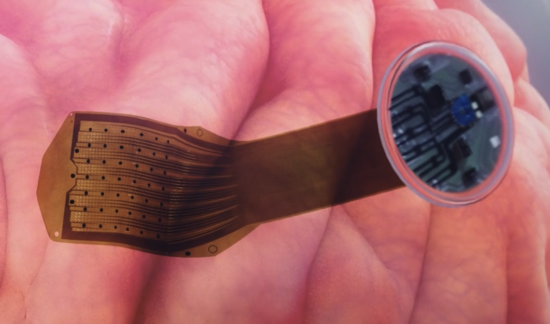
Implantation force: Russia has made neuroimplant technology safer
iz.ru
Nov. 10, 2026, 11:22 p.m.
Russian scientists from Sirius University of Science and Technology's Biomaterials department have developed and patented a novel material for soft electrodes that can stretch approximately 50% while maintaining electrical conductivity. This advancement addresses a critical limitation of conventional rigid electrodes used in neuroprostheses—devices designed to restore mobility and sensation following severe brain and spinal cord injuries. Rigid electrodes cause tissue damage, triggering inflammatory responses that create glial scars around implants, degrading signal quality within one to three months. The new electrically conductive two-layer nanocomposite coating on a flexible substrate mimics biological tissue properties, reducing mechanical irritation during brain movements and extending implant longevity. By minimizing tissue trauma, the soft electrodes could significantly improve neuroprosthetic performance for patients with paralysis or sensory loss. However, scientists emphasize that comprehensive clinical testing remains necessary before implementation. This innovation represents meaningful progress toward more effective neural restoration therapies for individuals with neurodegenerative conditions and catastrophic nervous system injuries.
Share on

Adapters & Interface Boards
www.neuronexus.com
Nov. 10, 2026, 11:22 p.m.
NeuroNexus offers a comprehensive solution for connecting incompatible probe packages and acquisition systems through connector-pair adapters, electrode interface boards, and breakout adapters. The primary product, the NeuroNexus Connector-Pair Adapter, provides 78 different pairings covering various input connectors including Omnetics, A-series, ZIF, CQ4, Q4, and QIB formats on the package side, matched against headstage outputs from leading manufacturers including Intan, Blackrock, A-M Systems, BrainAmp, and Plexon. Beyond basic adapters, NeuroNexus supplies electrode interface boards for EIB rails and X3/X3SR headstages, plus specialized breakout adapters such as the High-Density Matrix adapter and X-Series connector adapter for diverse experimental configurations. Remap adapters allow channel remapping, requiring verification of site-to-channel maps before recording. These solutions enable seamless integration across multiple neurophysiology platforms and connector standards, reducing compatibility barriers for researchers using varied equipment combinations.
TDT Neuroscience Tools & Software Solutions
www.tdt.com
Nov. 10, 2026, 11:22 p.m.
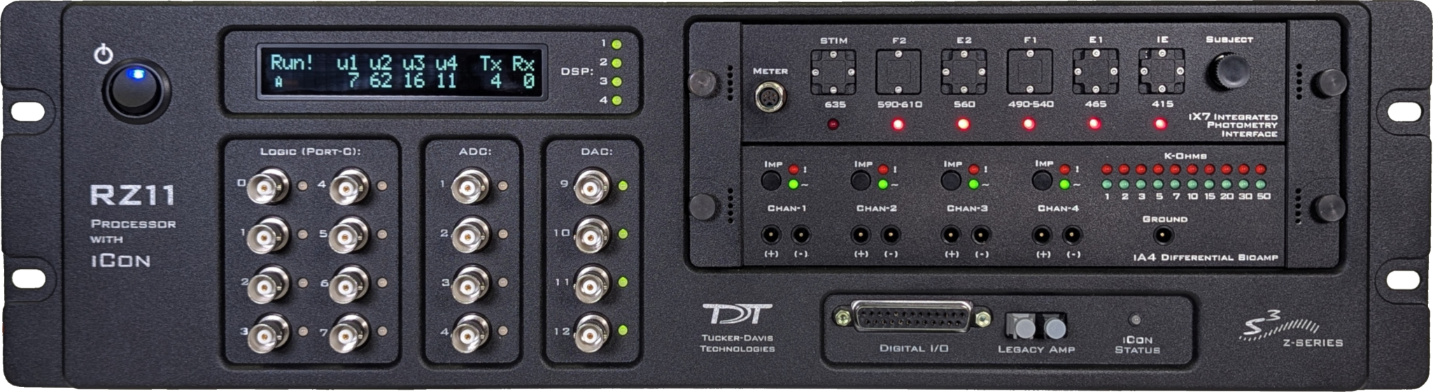
TDT systems have become instrumental in neuroscience research, with over 6,400 published studies utilizing these tools across diverse research areas and years. Recent applications demonstrate the platform's critical role in understanding neonatal brain injury. One study published in Clinical and Experimental Pediatrics examined how maternal metformin administration during pregnancy protects offspring from hypoxia-ischemia-induced auditory dysfunction. Using auditory brainstem responses (ABRs) and immunohistochemistry, researchers found that metformin exposure prevented HI-induced hearing threshold elevation and preserved neuronal density in the medial nucleus of the trapezoid body (MNTB). Additionally, a Materials Today Bio publication addressed cisplatin-induced ototoxicity, a significant clinical challenge causing hearing loss in approximately fifty percent of treated patients. The research explores sodium thiosulfate as an otoprotectant, though achieving sufficient cochlear concentrations remains problematic due to the organ's poor drug clearance. These studies highlight TDT's value in investigating sensory system pathology and evaluating therapeutic interventions for preventing irreversible hearing loss and neurological damage.
Share on
Injectable Mesh Electronics Could Enable Long-Term Neural Recording Without Scar Tissue Buildup
bioletric.com
Nov. 10, 2026, 11:21 p.m.
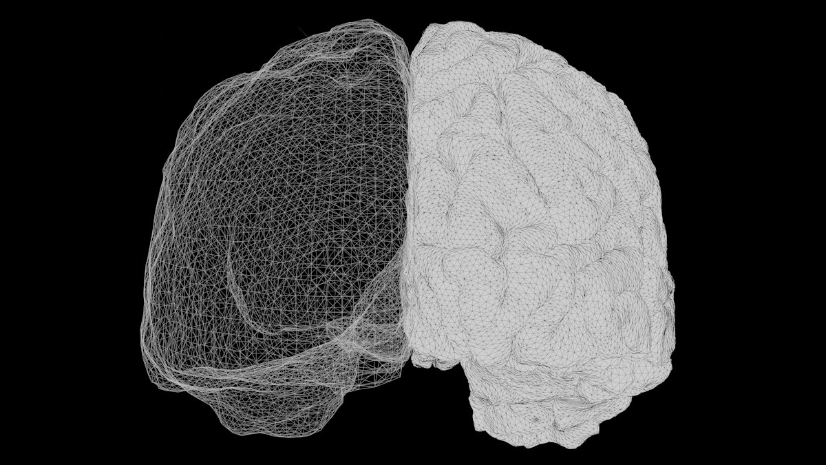
Researchers at Harvard, led by chemist Charles Lieber, have developed injectable mesh electronics designed to overcome a fundamental limitation in long-term brain-computer interfaces. Traditional rigid silicon electrode arrays, successfully demonstrated in landmark studies like the 2012 BrainGate trial where paralyzed patient Cathy Hutchinson controlled a robotic arm, suffer from a critical mechanical mismatch. Silicon's stiffness measured in gigapascals vastly exceeds brain tissue's kilopascal-range softness, causing chronic friction. This triggers an immune response where microglia and astrocytes form glial scar tissue around implants, insulating electrodes and degrading signal quality within months—a well-documented challenge across the field, including research at the University of Michigan. Rather than pursuing smaller probes with higher electrode counts, Lieber's team fundamentally reconceived neural electrodes to match brain tissue's mechanical properties. This approach, published in Nature, promises to maintain signal integrity in chronic applications, potentially enabling durable brain-computer interfaces for paralysis and neurological conditions.
Share on
Neural Probes: What They Are, How They Work, and How to Choose One
www.neuronexus.com
Nov. 10, 2026, 10:48 p.m.
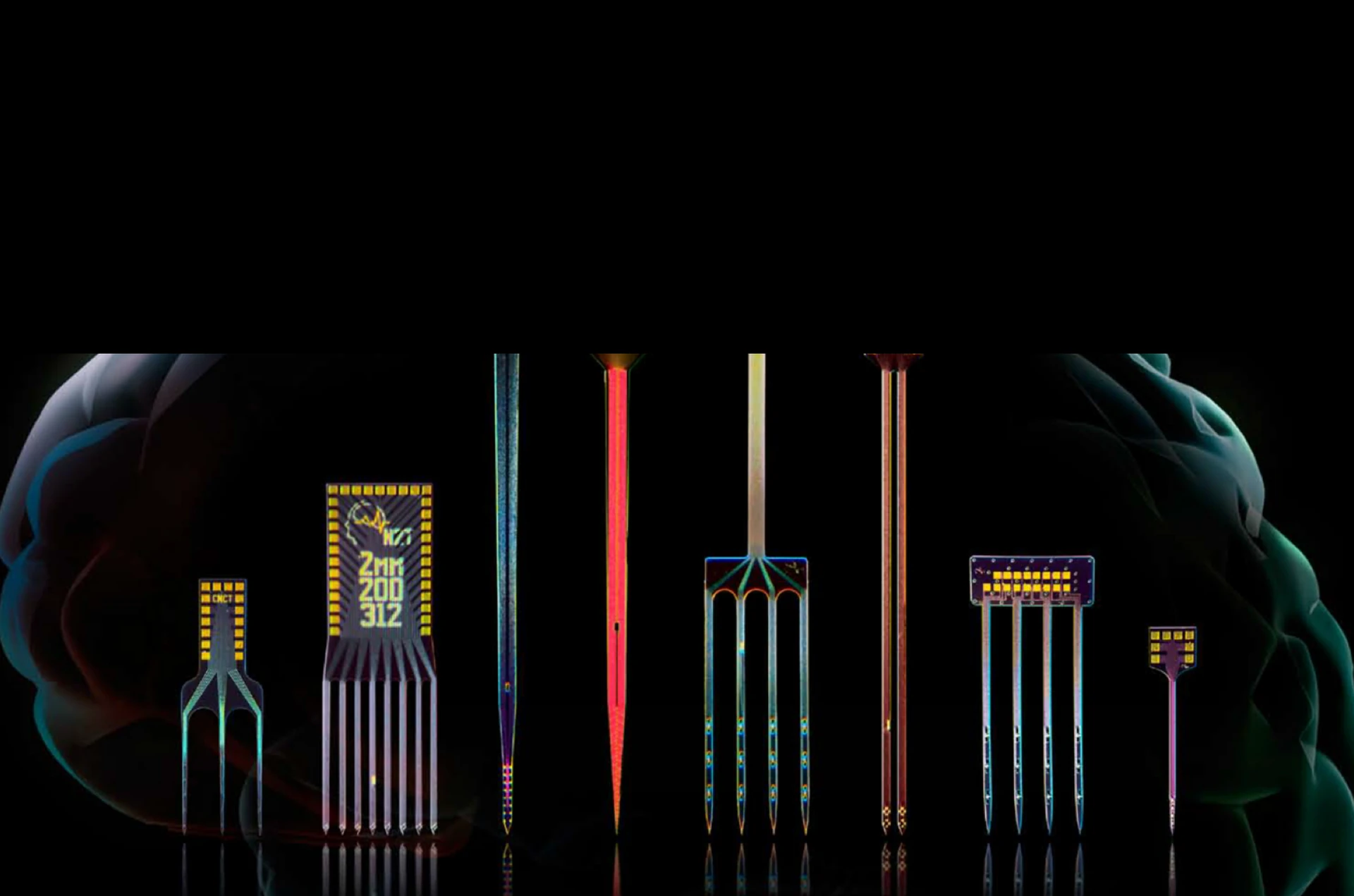
Neural probes are implantable microscale devices equipped with discrete electrode sites that record or stimulate neural activity in the brain, spinal cord, or peripheral nerves. Developed by pioneers like the University of Michigan group that created the foundational Michigan probe, these devices feature penetrating shanks ranging from 15–50 micrometers thick with one to over a thousand individually addressable electrode sites. While most probes are designed for electrical recording of extracellular biopotentials—capturing action potentials and local field potentials—modern neural probes offer multimodal capabilities including optical and chemical sensing through integrated micro-LEDs, optical fibers, and microfluidic channels. The devices can safely stimulate neural structures using charge-balanced current pulses of 10–200 microamperes. NeuroNexus, a leading provider in this space, offers a comprehensive catalog spanning probe designs from single to eight-shank configurations. Silicon-substrate probes are particularly prevalent due to their precision and scalability. These technologies enable both acute experimental applications and chronic clinical implants, supporting advanced neuroscience research and therapeutic interventions across various neurological conditions.
Share on
XDAQ CORE2 | NeuroNexus
www.neuronexus.com
Nov. 10, 2026, 10:48 p.m.
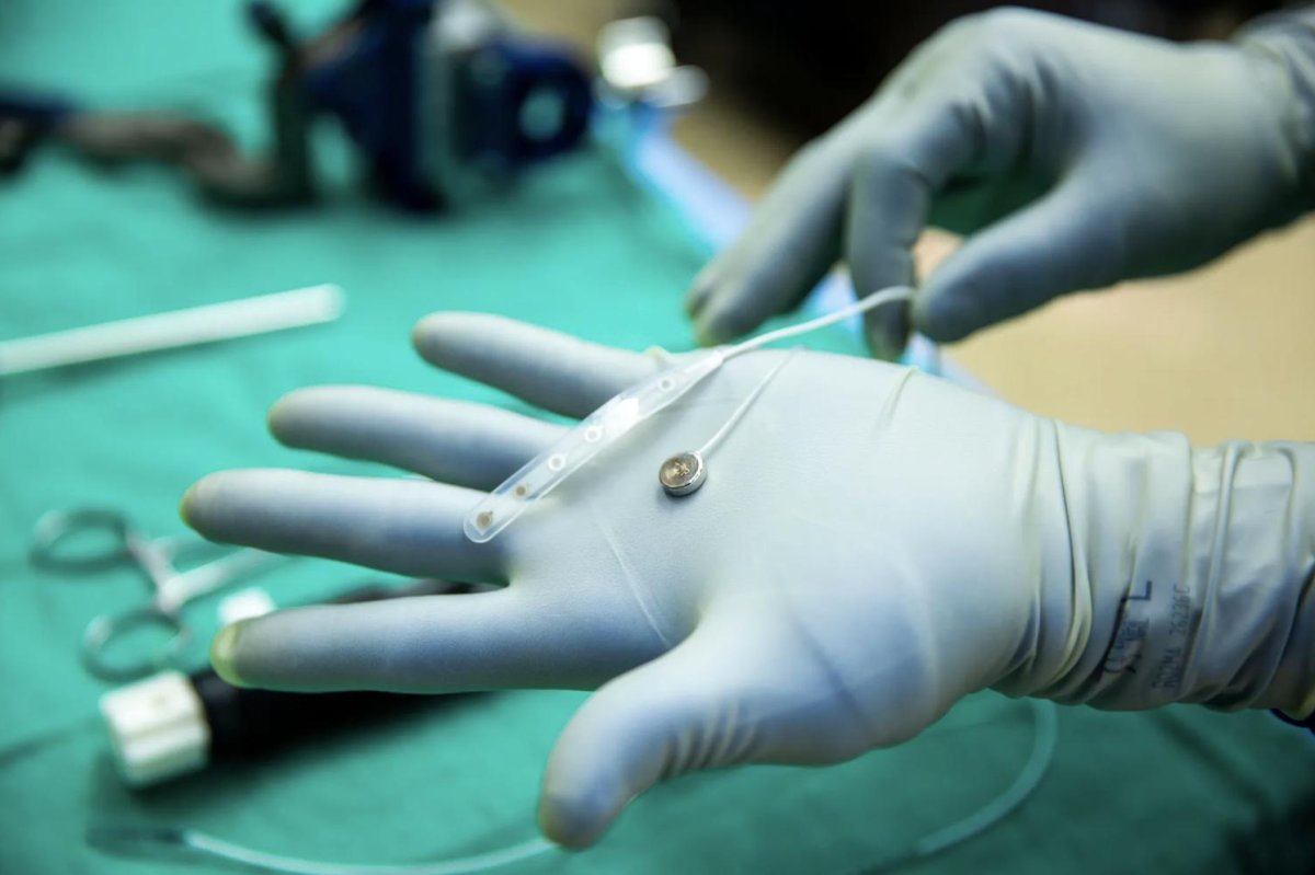
The XDAQ CORE2 represents an advanced 512-channel acquisition system designed for high-density in vivo electrophysiology research. Operating at 30 kS/s with 16-bit resolution, the system supports simultaneous recording and stimulation across up to 512 channels via Thunderbolt 3+ connectivity. Compatible with industry-standard Intan RHD/RHS headstages and Neuropixels probes, the XDAQ CORE2 enables researchers to capture neural activity with minimal latency while maintaining integrated synchronization across channels. This capability is particularly valuable for complex neuroscience experiments requiring simultaneous multi-site recordings. The platform offers configurable options allowing users to tailor specifications to specific experimental requirements. Researchers can customize their setup, obtain detailed pricing and lead-time information, and receive comprehensive usage guidance within one business day. The system has already been cited in multiple peer-reviewed publications, establishing its relevance in contemporary neuroscience research and supporting the growing demand for high-channel-count electrophysiological recording solutions.
Share on
Silicon Neural Probes and Thin-Film Grids: An Introduction
www.neuronexus.com
Nov. 10, 2026, 10:48 p.m.
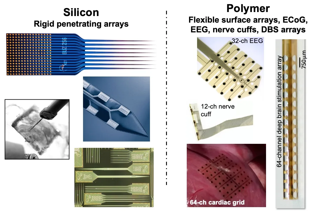
NeuroNexus has introduced a comprehensive overview of two complementary thin-film electrode platforms for neural and cardiac electrophysiology research. Silicon probes are rigid, penetrating arrays designed to record and stimulate activity at precisely targeted sites within brain tissue, spinal cord, nerves, ganglia, or heart, while thin-film polymer grids are flexible surface arrays that non-invasively measure electrical activity beneath cortical, cardiac, or nerve surfaces. Both platforms leverage proprietary photolithography and microfabrication techniques to ensure consistent, high-fidelity electrode reproduction. With over twenty years of innovation building upon the foundational Michigan probe design, NeuroNexus provides researchers with complementary solutions for direct electrophysiological measurements across relevant spatial and temporal scales. This dual-platform approach addresses diverse research applications, from mainstream studies to leading-edge neuroscience and cardiac investigations, supported by integrated instrumentation and analytical software for reliable signal acquisition and data management.
Share on

Types of BCI: Invasive, Semi-invasive, and Non-invasive—Possibilities and Limitations|Tex
note.com
Nov. 10, 2026, 10:47 p.m.
Brain-computer interfaces represent a diverse technological landscape fundamentally shaped by electrode placement within the brain. Three primary categories exist, each with distinct characteristics. Invasive BCIs position electrodes directly into brain tissue using silicon arrays like the Utah Array or flexible thread electrodes similar to Neuralink's design, requiring craniotomy but delivering the highest signal quality capable of recording individual neuron activity from hundreds to thousands of neurons simultaneously. Semi-invasive electroencephalography on the cortex (ECoG) places electrode grids on the brain surface without penetrating tissue, offering a middle ground between invasive and non-invasive approaches with significantly higher signal quality than scalp-based methods yet lower surgical risks. Endovascular systems like Synchron's technology navigate brain blood vessels via catheter surgery, providing medium to high signal quality with reduced invasiveness. Non-invasive electroencephalography requires no surgery but operates with lower signal quality. Recent breakthroughs demonstrate invasive BCIs enabling remarkable capabilities: BrainGate's spinal cord injury patients controlling robotic arms through thought alone, Neuralink's quadriplegic user operating computer cursors and playing chess, and Stanford's speech BCI achieving 90 characters-per-minute input. Despite risks including infection and electrode degradation, clinical trials are advancing, with FDA medical device approval anticipated in the late 2020s to early 2030s for specific indications like ALS.
Share on
The brain implant threaded up a neck vein, no skull opening: Synchron's Stentrode endovascular BCI
www.kpsphere.com
Nov. 10, 2026, 10:46 p.m.
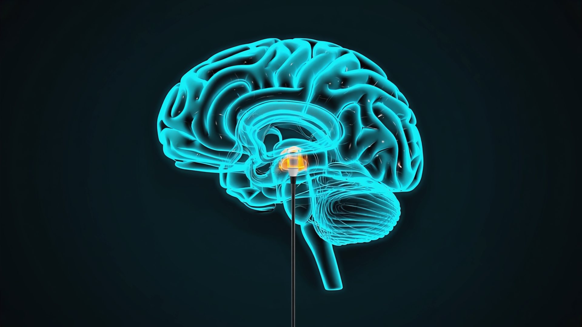
Researchers have developed an innovative brain implant that can be inserted through a neck vein, eliminating the need for invasive skull surgery. This minimally invasive approach represents a significant advancement in neural interface technology, as traditional brain implants have required cranial procedures that carry substantial surgical risks and complications. The endovascular technique threads the device up through the venous system to reach the brain, substantially reducing patient trauma and recovery time. This breakthrough is particularly significant for patients who require brain monitoring or therapeutic intervention but face elevated surgical risks. The development combines AI-assisted research methodologies with advanced medical device engineering to achieve a safer alternative to conventional neurosurgical implantation. This technique could substantially expand access to brain implant therapies by making the procedure more accessible to broader patient populations and reducing associated healthcare burdens. The innovation demonstrates how emerging technologies can address critical limitations in existing medical procedures, potentially transforming treatment options for neurological conditions.
Share on
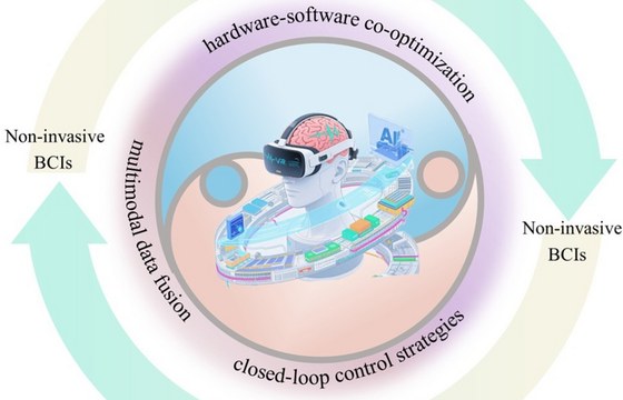
Intelligent Biosensing for Brain-computer Interfaces: From Neural Signal Acquisition to AI-powered Decoding
www.intechopen.com
Nov. 7, 2026, 5:17 a.m.
Brain-computer interfaces (BCIs) establish direct communication pathways between the brain and external devices, unlocking new possibilities for understanding and harnessing human neural capabilities. This comprehensive review examines current progress in intelligent sensing technology for BCIs, encompassing neural signal acquisition methods and AI-powered decoding approaches. The analysis traces BCIs' evolutionary development, system architecture, and research importance, then evaluates multimodal neural signal sensing technologies spanning from non-invasive to invasive modalities, assessing their respective performance characteristics. The review emphasizes artificial intelligence's critical role in neural signal decoding, particularly deep learning applications ranging from traditional machine learning to advanced architectures including Transformers and large language models, alongside multimodal fusion and edge computing optimization techniques. Practical BCI applications in medical rehabilitation, neuro-ergonomics, and intelligent interaction are examined, with discussion of technical challenges and ethical considerations. As neural signal acquisition methods and AI decoding algorithms continue advancing, intelligent BCI sensing technology promises transformative impacts across medical rehabilitation, human-computer interaction expansion, and human potential enhancement, driving significant societal change.
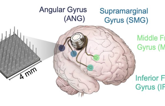
Long-term stability of cellular-resolution brain-computer
simonjacob.de
Nov. 7, 2026, 5:17 a.m.
Researchers have demonstrated the long-term stability and viability of cellular-resolution brain-computer interface recordings in stroke patients. This study addresses a critical challenge in BCIs: maintaining high-fidelity neural signal detection over extended periods following brain injury. The investigation evaluated how cellular-resolution recording systems perform in post-stroke conditions, where neural tissue undergoes significant changes including inflammation and reorganization. The findings indicate that these advanced recording interfaces can sustain reliable, single-neuron-level signal acquisition despite the hostile post-stroke neurological environment. This stability is crucial for developing practical BCIs for stroke rehabilitation and motor recovery applications, as it suggests that implanted electrode arrays can maintain their sensitivity and recording quality without requiring frequent replacement or recalibration. The results validate the potential of cellular-resolution BCIs as long-term clinical tools, offering hope for stroke patients seeking to regain motor function through brain-computer interface technology and establishing a foundation for future therapeutic applications in neurorehabilitation.
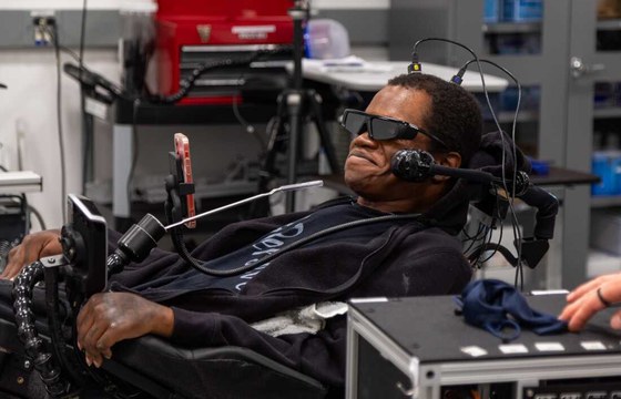
Industry News Briefs from the International Neuromodulation Society
www.google.com
Nov. 7, 2026, 5:16 a.m.
Recent breakthroughs in neuromodulation and brain-computer interfaces are expanding therapeutic possibilities across multiple neurological and psychiatric conditions. A landmark achievement by researchers at UC San Francisco and UC Berkeley demonstrates an AI-powered brain implant that successfully restored speech to Ann Johnson, a stroke survivor paralyzed for nearly two decades. The implant decodes her brain signals to generate speech and facial expressions through a computer avatar. Simultaneously, emerging therapies are gaining clinical momentum: transcranial magnetic stimulation (TMS) shows promise for depression, OCD, and addiction disorders, though accessibility challenges persist; vagus nerve stimulation demonstrates therapeutic potential for depression, obesity, and inflammatory conditions through techniques like cold exposure and controlled breathing; and deep brain stimulation during specific sleep phases enhances memory consolidation in epilepsy patients, with implications for Alzheimer's disease treatment. A clinical review addresses the safety and effectiveness of neuromodulation techniques including vagal nerve stimulation and responsive neurostimulation during pregnancy and lactation. These advances represent significant progress in treating previously intractable neurological and psychiatric conditions, though implementation challenges surrounding clinic accessibility and insurance coverage remain important considerations.
Share on
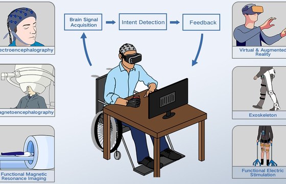
Brain-Computer Interface (BCI) for Stroke Rehabilitation | Sağlık Rehberi
saygi.tr
Nov. 7, 2026, 5:16 a.m.
Brain-computer interfaces (BCIs) represent a promising approach to stroke rehabilitation by translating cortical electrical activity—typically recorded via EEG—into control commands for external devices such as robotic exoskeletons, functional electrical stimulation (FES) systems, virtual reality avatars, or assistive robots. The technology works by detecting sensorimotor rhythm changes when patients imagine moving their paretic limb, thereby harnessing neuroplasticity and closing the feedback loop between motor intention and proprioceptive or visual feedback. Motor imagery-based BCI protocols have demonstrated improved clinical outcomes, including better Fugl-Meyer and ARAT scores, particularly in chronic stroke patients with severe upper-limb paresis compared to sham interventions. Successful implementation demands multidisciplinary collaboration among neurologists, biomedical engineers, and rehabilitation specialists, alongside careful patient selection based on cognitive ability and scalp accessibility. Current research is advancing toward hybrid EEG-EMG systems, implantable electrode arrays for severe paralysis, home-based BCI applications, and integration with transcranial stimulation techniques. This evolving field offers significant potential to enhance motor recovery and functional independence in stroke survivors.
NMPA Clinical Trial Guideline on Implanted BCI Hand-Movement Compensation System: Key Focus on Patient Eligibility, Clinical Endpoints, and Lifecycle Evidence
chinameddevice.com
Nov. 7, 2026, 5:16 a.m.
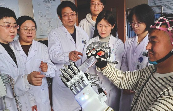
China's National Medical Products Administration released its final clinical trial guideline for implanted brain-computer interface hand-movement compensation systems on October 22, 2026, building on the FDA's framework for BCI devices. The guideline mandates clinical trials to confirm integrated safety and effectiveness, with overseas data potentially acceptable as sufficient evidence. For feasibility assessment, at least four patients with the same indication are recommended. Where no effective alternative treatment exists and no neural repair is claimed, single-arm designs with objective performance goals are permitted. The primary endpoint measures six-month BCI-assisted ARAT grasping response rates, with a minimum clinically important difference of approximately two points and sample sizes of at least 32 patients justified statistically. Follow-up must extend at least six months, with instructions for use specifying calibration thresholds and MRI compatibility. Post-market surveillance requires minimum two-year data collection on signal stability, decoding accuracy, performance degradation, mental burden, and safety metrics. The guideline specifically targets implanted epidural systems with local field potential decoding using 64 or fewer channels, designed for hand grasping via pneumatic gloves in tetraplegia patients with C2–C6 cervical spinal cord injuries aged 18–60 years, excluding non-implanted alternatives and neural repair claims.
Share on
Headstage — the Digitizing Preamplifier at the Head
www.neuronexus.com
Nov. 3, 2026, 12:58 p.m.
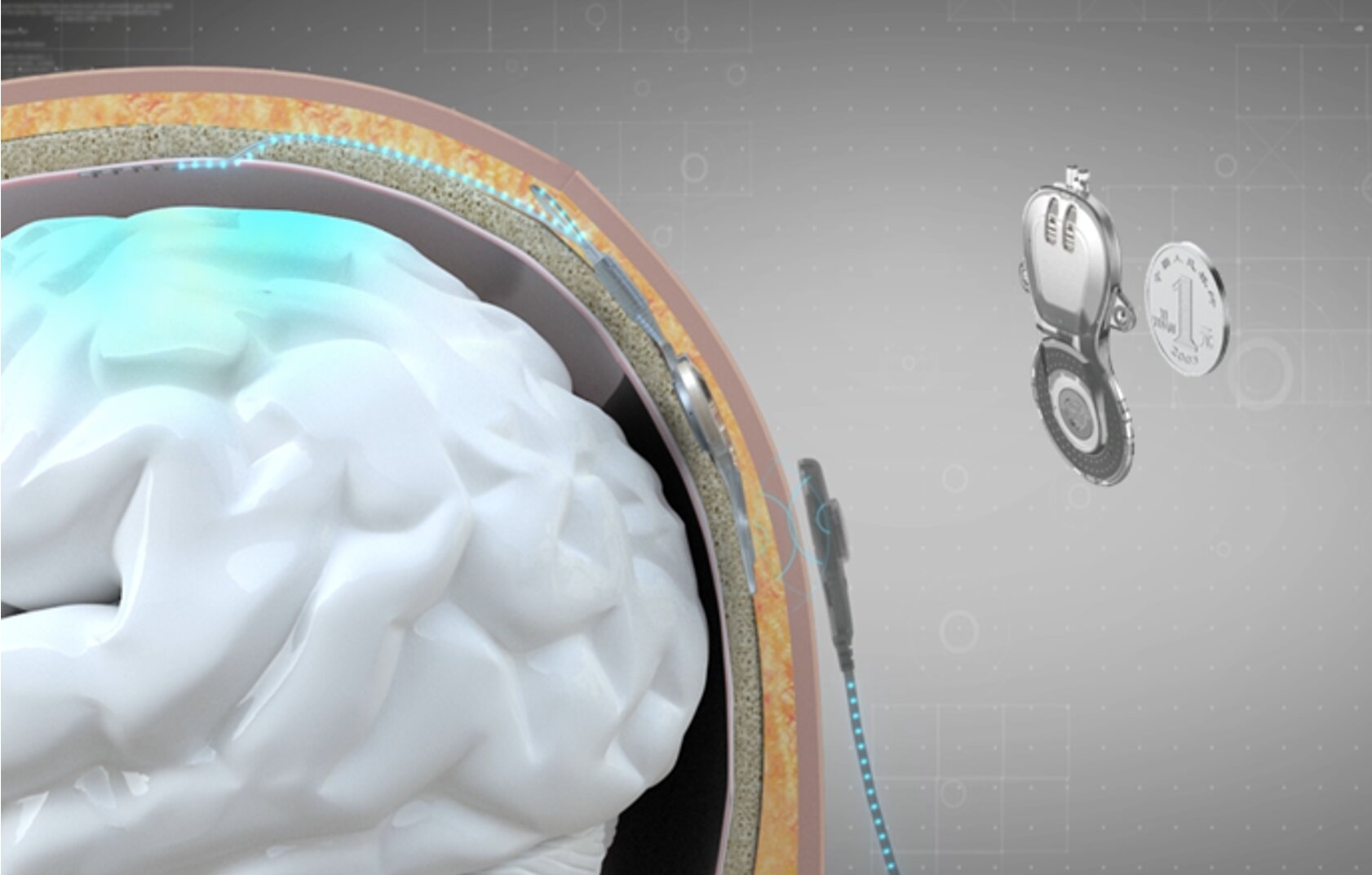
NeuroNexus manufactures headstages—preamplifiers positioned at the recording probe to digitize neural signals at the source. These devices integrate Intan RHD chips for recording or RHS chips for simultaneous recording and stimulation, combining amplifiers, programmable filters, and 16-bit analog-to-digital converters onto a single die. The technology achieves input-referred noise of approximately 2.4 µV rms with sampling rates up to 30 kS/s and software-configurable pass-bands spanning below 1 Hz for field potentials to several kilohertz for spike detection. By digitizing signals near the probe tip, the system minimizes analog cable length to millimeters, maintaining low noise floors while requiring only a single micro-HDMI or SPI link regardless of channel count—whether 16 or 128 channels. Headstages also measure electrode site impedance in situ and can synchronize accelerometer data with neural recordings. NeuroNexus offers two product lines: X-Series headstages, engineered by Kontex, weighing 0.9–1.5 grams in record-only or stimulation-capable configurations; and SmartLink headstages using various connector types for acute and bench applications. This architecture addresses fundamental noise and scalability challenges in multi-channel neural recording systems.
Share on
Editorial: BCIs for monitoring and treating mental health conditions
www.frontiersin.org
Nov. 3, 2026, 12:58 p.m.
Brain-computer interfaces (BCIs) offer significant promise for monitoring and treating mental health conditions, yet clinical adoption remains limited due to low awareness among healthcare providers. Researchers from the Technical University Dresden and National University of Singapore have surveyed available BCI technologies spanning non-invasive methods like EEG and functional near-infrared spectroscopy to invasive approaches including epicortical electrocorticography and intracortically implanted electrodes. A notable study by Belkacem et al. employed EEG with deep residual multilayer perceptron computational modelling to demonstrate that music therapy modulates brain activity patterns, improving discrimination of mental fatigue states. Building on established applications in Parkinson's disease, where reduced beta-band power correlates with motor impairment, researchers have expanded BCI applications to stroke rehabilitation. Su et al. developed a protocol investigating sensorimotor beta-band activity dynamics combined with action observation and object affordance training to enhance motor cortical excitability and visuomotor processing during motor imagery, aiming to improve BCI performance and upper-limb functional recovery. These advances suggest EEG-based biomarkers could be integrated into routine clinical practice for personalized treatment, improved therapeutic outcomes, and better prediction of clinical trajectories in psychiatric and neurological disorders.
Share on
EEG-guided robotic control of transcranial magnetic
www.biorxiv.org
Nov. 3, 2026, 12:58 p.m.
# EEG-Guided Robotic Control of Transcranial Magnetic Stimulation
Researchers have developed an innovative system that integrates electroencephalography (EEG) with robotic technology to enhance the precision and efficacy of transcranial magnetic stimulation (TMS). This advancement represents a significant breakthrough in neuromodulation therapy, particularly for treating neuropsychiatric conditions such as depression and chronic pain.
The system employs real-time EEG monitoring to guide robotic positioning of TMS coils, enabling targeted stimulation of specific brain regions based on individual neural activity patterns. By combining non-invasive brain imaging with automated coil placement, the technology achieves superior spatial accuracy compared to conventional manual TMS administration. This EEG-guided robotic approach allows clinicians to identify optimal stimulation parameters tailored to each patient's unique neurophysiological profile.
The development of this integrated technology matters considerably because it potentially improves treatment outcomes by maximizing therapeutic effectiveness while minimizing off-target effects. The system's ability to adapt stimulation protocols based on real-time brain activity monitoring could transform TMS from a one-size-fits-all approach into a truly personalized intervention. This innovation has important implications for advancing precision medicine in neurology and psychiatry, offering patients more effective and safer therapeutic options.
Share on
EEG datasets of stroke patients (motor imagery)
nemar.org
Nov. 3, 2026, 12:58 p.m.
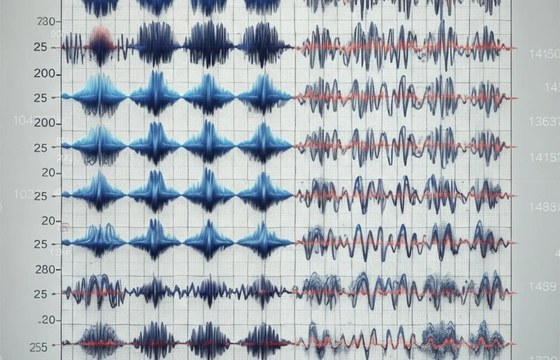
This dataset, curated by Liu, Lv and colleagues and published in 2023, comprises electroencephalography recordings from 50 stroke patients engaged in motor imagery and imagery tasks. The collection totals 1.15 GB and is formatted according to HED version 8.4 standards. The data is openly accessible through NEMAR (a neuroimaging data repository) under a Creative Commons Attribution 4.0 license, enabling broad use for research purposes. Multiple access methods are provided, including direct download via the NEMAR command-line interface, Zarr-based cloud access for efficient subset retrieval, and HTTPS protocols for individual files. The dataset includes participant demographics and per-file data quality assessments generated through the NEMAR processing pipeline. This resource is valuable for stroke rehabilitation research, brain-computer interface development, and motor recovery studies, as motor imagery paradigms are critical for understanding neural plasticity following stroke and informing neurorehabilitation interventions. The open-access approach facilitates reproducible research and cross-institutional collaboration in translational neuroscience.
Brain-Computer Interface For Stroke Rehabilitation: How EEG & FES Work
www.fastturnpcbs.com
Nov. 3, 2026, 12:58 p.m.
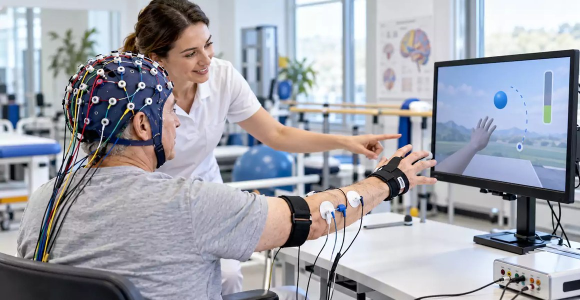
Brain-computer interfaces (BCIs) represent an innovative approach to stroke rehabilitation by detecting movement intentions through electroencephalography (EEG) and converting them into functional feedback mechanisms. These systems translate brain activity into outputs such as functional electrical stimulation (FES), robotic assistance, or visual cues, enabling patients with severe hand and arm impairment to begin rehabilitation training through attempted or imagined movements even when physical movement is compromised. By combining real-time signal processing with sensorimotor feedback, BCI rehabilitation leverages neuroplasticity to support motor recovery through repeated training cycles. The technology addresses a critical clinical need: traditional rehabilitation requires voluntary movement that severely impaired stroke survivors often cannot generate. EEG-based BCIs measure electrical activity via scalp electrodes positioned over sensorimotor regions, typically at locations C3 and C4 for hand rehabilitation. Signals pass through an analog front end for amplification and conditioning before digital conversion. System effectiveness depends on clinical factors including impairment severity, signal quality, training protocols, and individual patient characteristics. BCI rehabilitation shows promise for both acute stroke patients and those in chronic phases experiencing persistent movement limitations months or years post-stroke.
Share on